How To Read Sonogram
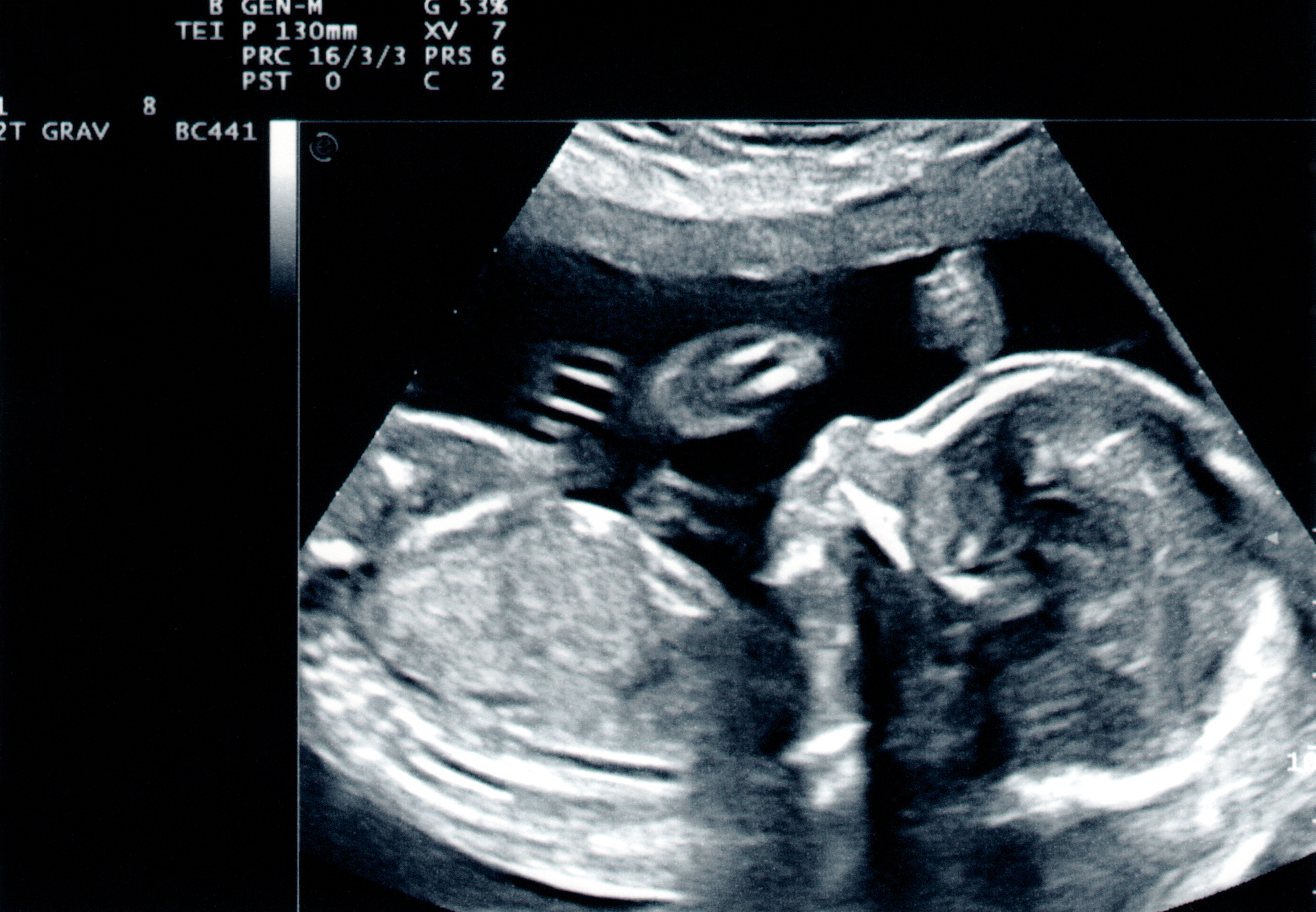
How To Read Sonogram - The technology behind the difference between ultrasound and sonogram. Sensors attached to the chest and sometimes the legs check the heart. Lean about the various sections of report including type of exam, history/reason for exam, comparison/priors, technique, findings,. Depending on the part of the body that you’re looking at, you may need to find the walls of the uterus or the. The radiologist writes the report for your provider who ordered the exam. Here you can see the organs or tissues. Here’s a brief explanation of how to read ultrasound numbers and what they mean: Orientation you have to determine the orientation of. The rig includes openings into which the ultrasound module can be affixed, with. An echocardiogram uses sound waves to show how blood flows through the heart and heart valves.
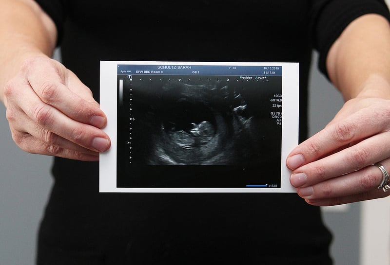
Web however, there’s a difference between the two: The water balloon of the transducer was coupled to the mouse head using different coupling methods, as shown in fig. Ultrasound centers and hospitals tend to use this space for details such as: The rig includes openings into which the ultrasound module can be affixed, with. For instance, at the top of the ultrasound images of your. Web sonogram definition, the visual image produced by reflected sound waves in a diagnostic ultrasound examination. Ultrasound examination is less expensive to perform than ct or mri. An ultrasound picture is called a sonogram. You can also see ultrasound numbers when obtaining a fetal image, aside from the image itself. A sonogram is the picture that the ultrasound generates.
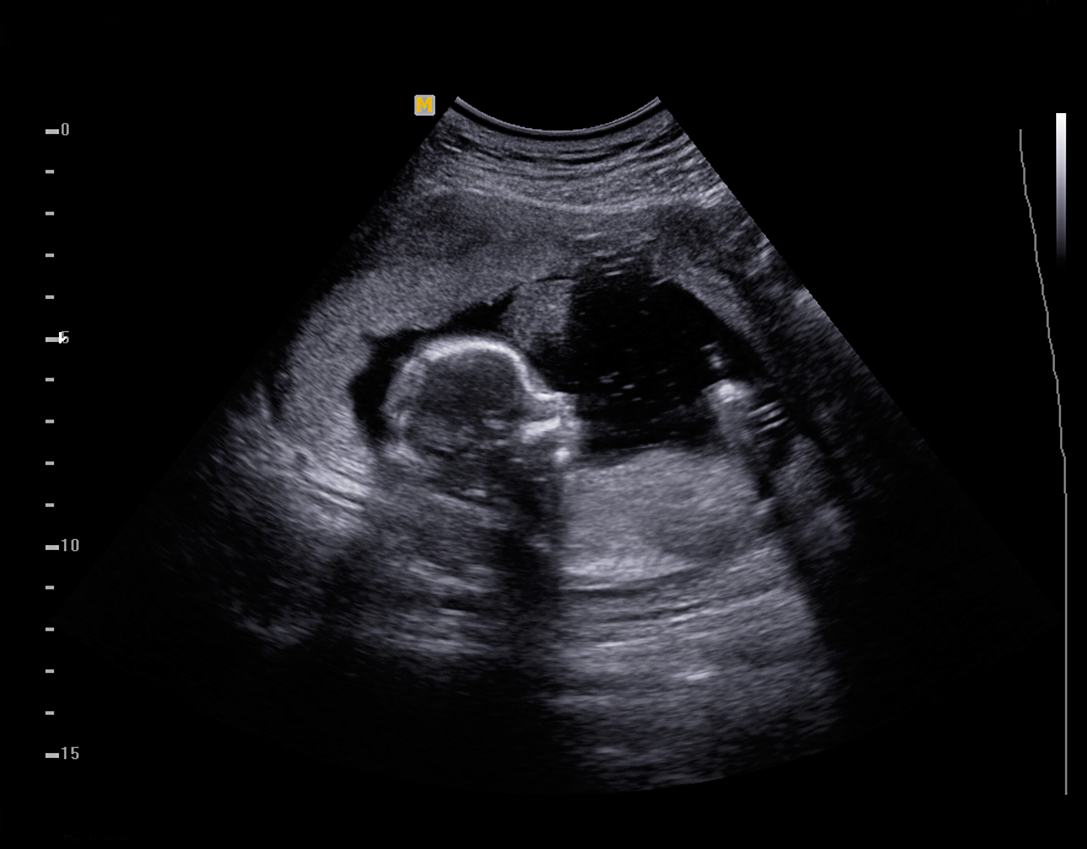
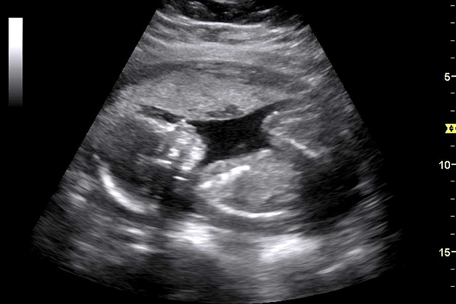
Ultrasound (us) use has rapidly entered the field of acute pain medicine and regional anesthesia and interventional pain medicine over the last decade, and it may even become the standard of practice. Web begin from the top now, you look at the top of the image. The size and shape of your. Ultrasound examination is less expensive to perform than ct or mri. Here you can see the organs or tissues. Ultrasound centers and hospitals tend to use this space for details such as: Lean about the various sections of report including type of exam, history/reason for exam, comparison/priors, technique, findings,. Web acoustic coupling methods. Web sonogram definition, the visual image produced by reflected sound waves in a diagnostic ultrasound examination. Web the system consists of a piezoelectric ultrasound scanning module that fits into a rig that can be affixed to a bra.

estatenygw pregnancy week 6 ultrasound photos
Web an echo test can allow your health care team to look at your heart’s structure and check how well your heart functions. The ultrasound is the process to retrieve the information and the sonogram is the end picture showing the result. Web however, the best way to define the contract between sonogram vs ultrasound would be this: For instance,.

baby sonogram YouTube
Ultrasound centers and hospitals tend to use this space for details such as: Here’s a brief explanation of how to read ultrasound numbers and what they mean: Orientation you have to determine the orientation of. Ultrasound (us) use has rapidly entered the field of acute pain medicine and regional anesthesia and interventional pain medicine over the last decade, and it.
OneCall24
Orientation you have to determine the orientation of. Web ultrasound (also called sonography or ultrasonography) is a noninvasive imaging test. Ultrasound (us) use has rapidly entered the field of acute pain medicine and regional anesthesia and interventional pain medicine over the last decade, and it may even become the standard of practice. An ultrasound picture is called a sonogram. Lean.

6 Ways to Tell Baby's Gender From an Early Sonogram
Depending on the part of the body that you’re looking at, you may need to find the walls of the uterus or the. The technology behind the difference between ultrasound and sonogram. Ultrasound (us) use has rapidly entered the field of acute pain medicine and regional anesthesia and interventional pain medicine over the last decade, and it may even become.
Sonogram vs Ultrasound A More InDepth Distinction Between The Two
Ultrasound centers and hospitals tend to use this space for details such as: Web however, there’s a difference between the two: Typically, the radiologist sends the report to the person who ordered your test, who then delivers the results to. Color an ultrasound or sonogram picture is a black and white photograph, so they all look the same to someone.
First Look at Your Baby The Fascinating History of the "Sonogram"
Web however, the best way to define the contract between sonogram vs ultrasound would be this: The rig includes openings into which the ultrasound module can be affixed, with. Web information to help patients understand their abdominal ultrasound radiology report. An ultrasound is a tool used to take a picture. Lean about the various sections of report including type of.

Baby sonogram ornament please read full description before Etsy
Here’s a brief explanation of how to read ultrasound numbers and what they mean: For the “oil + hairs” group, mineral. It shows where the probe was placed during the ultrasound. Here you can see the organs or tissues. The radiologist writes the report for your provider who ordered the exam.

Sonogram showing baby giving thumbs up in the womb goes viral ABC11
Here’s a brief explanation of how to read ultrasound numbers and what they mean: Here you can see the organs or tissues. Ultrasound examination is less expensive to perform than ct or mri. It shows where the probe was placed during the ultrasound. You can also see ultrasound numbers when obtaining a fetal image, aside from the image itself.
How to Read an Ultrasound Gender and And Abnormality? New Health Advisor
Web the system consists of a piezoelectric ultrasound scanning module that fits into a rig that can be affixed to a bra. For the “oil + hairs” group, mineral. A sonogram is the picture that the ultrasound generates. Web acoustic coupling methods. An ultrasound is a tool used to take a picture.
Sonogram SG1 free sonogram by agworks
Ultrasound centers and hospitals tend to use this space for details such as: The test helps your health care team find out: For instance, at the top of the ultrasound images of your. Ultrasound (us) use has rapidly entered the field of acute pain medicine and regional anesthesia and interventional pain medicine over the last decade, and it may even.
Web Ultrasound (Also Called Sonography Or Ultrasonography) Is A Noninvasive Imaging Test.
Web however, the best way to define the contract between sonogram vs ultrasound would be this: Lean about the various sections of report including type of exam, history/reason for exam, comparison/priors, technique, findings,. An echocardiogram uses sound waves to show how blood flows through the heart and heart valves. The ultrasound is the process to retrieve the information and the sonogram is the end picture showing the result.
The Technology Behind The Difference Between Ultrasound And Sonogram.
Web begin from the top now, you look at the top of the image. Here you can see the organs or tissues. It shows where the probe was placed during the ultrasound. Ultrasound centers and hospitals tend to use this space for details such as:
Web An Echo Test Can Allow Your Health Care Team To Look At Your Heart’s Structure And Check How Well Your Heart Functions.
Web explaining the ultrasound numbers. An ultrasound picture is called a sonogram. You can also see ultrasound numbers when obtaining a fetal image, aside from the image itself. The size and shape of your.
The Water Balloon Of The Transducer Was Coupled To The Mouse Head Using Different Coupling Methods, As Shown In Fig.
Orientation you have to determine the orientation of. Web as you look at the ultrasound, you should try to locate all of the landmarks that you need to find. Ultrasound (us) use has rapidly entered the field of acute pain medicine and regional anesthesia and interventional pain medicine over the last decade, and it may even become the standard of practice. Typically, the radiologist sends the report to the person who ordered your test, who then delivers the results to.